
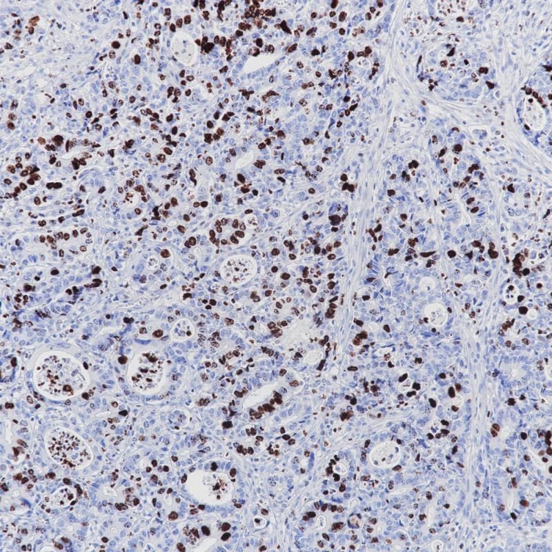
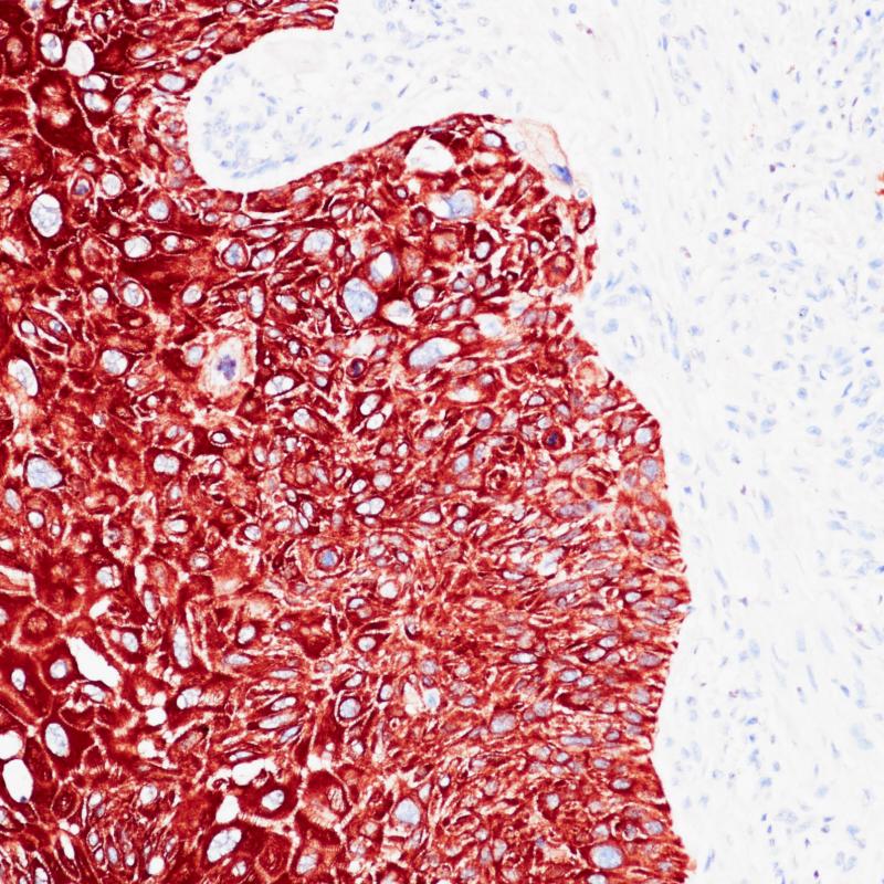
Gastric Carcinoma
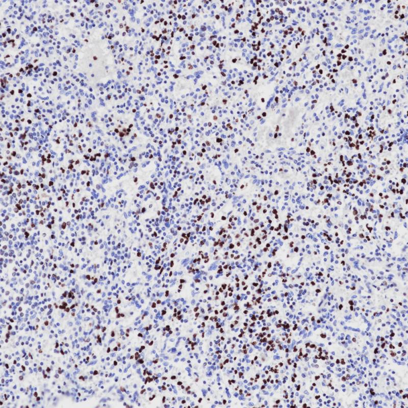
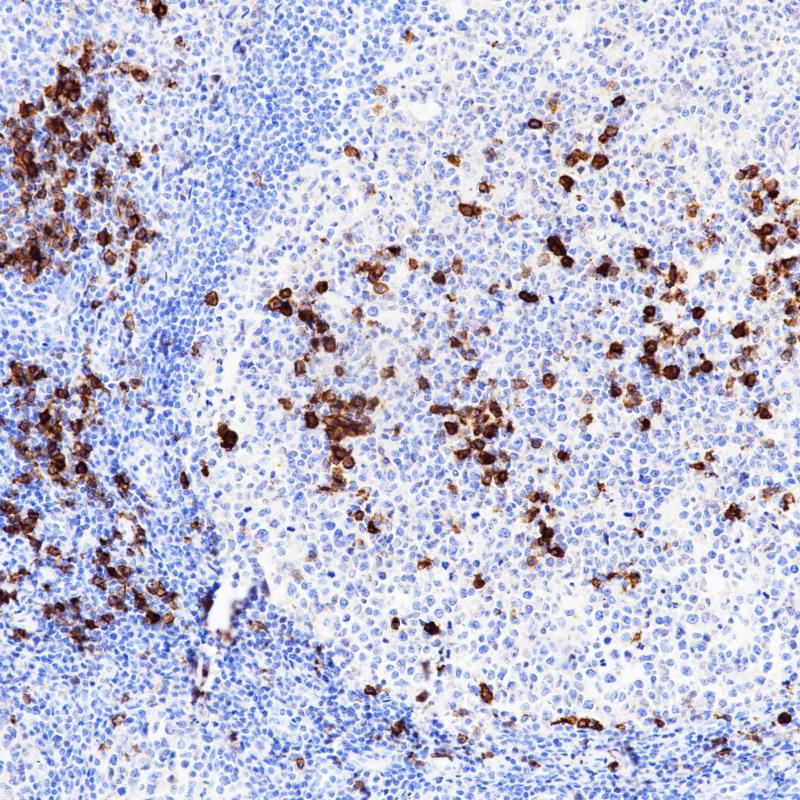
Spleen
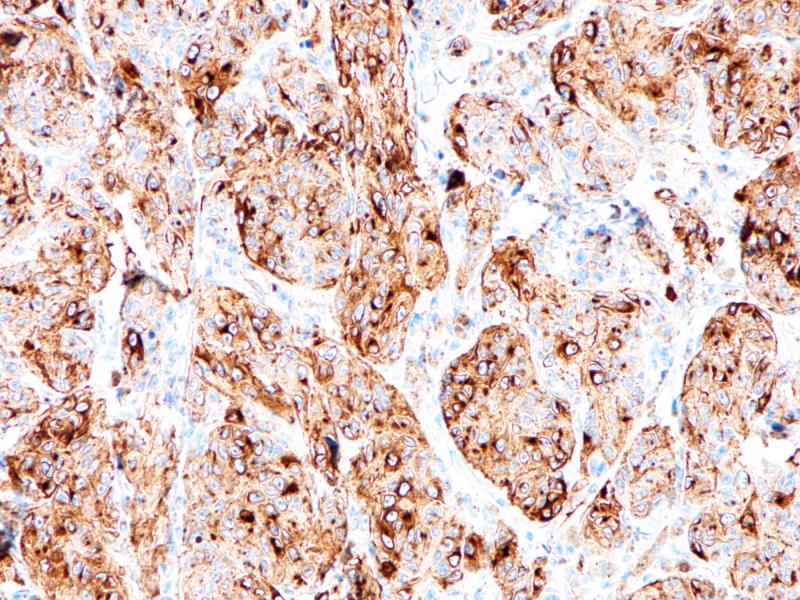
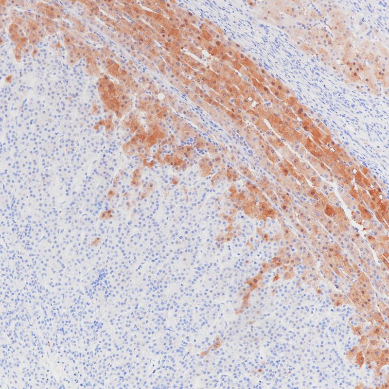
Related Products
Phosphohistone H3 (PHH3) Recombinant Rabbit Monoclonal Antibody
Phosphohistone H3 (PHH3) is a marker specific for cells undergoing mitosis. Serine 10 of Histone H3 is phosphorylated in association with mitotic chromatin condensation in late G2 and M phase of the cell cycle and thus, PHH3 can distinguish mitosis from apoptotic nuclei. The range of percentage PHH3 positive tumor nuclei was from 0.0 to 6.6% (median value 0.8%). Increased expression of PHH3 was significantly associated with tumor thickness (p = 0.031), presence of tumor ulceration (p =0.041) and tumor necrosis (p = 0.027), but not with Clark's level of invasion. High levels of PHH3 was associated with increased mitotic count (p = 0.003) and high Ki-67 expression (p = 0.002). For central nervous system tumors, melanoma, soft tissue tumors, GIST, etc., PHH3 mAb is helpful for tumor pathological classification and prognosis.
Specifications
- Catalog No.
- BX50087
- Clone No.
- BP6092
- Application
- IHC-P
- Subcellular location
- Nucleus
- Control
- Tonsil Tissue
- Recommended method
- HIER
- Volume
- 100μl/vial, 1ml/vial
- Dilution
- 1:100-1:200
- Immunogen
- Synthetic peptide corresponding to Phosphohistone H3 (PHH3) residues within aa1-100 (phospho S10) was used as an immunogen.
Reference
1.Thareja S, et al. Am J Dermatopathol. 2014 Jan; 36(1):64-7.
2.Casper DJ, et al. Am J Dermatopathol. 2010 Oct; 32(7):650-4.
Support Documents
Order
- E-mail : sales@biolynx.cn